
GI Efficacy Service Overview
Real patient tissue, three disease axes, and a clinical benchmark in every run
VivoSim Labs offers an integrated gastrointestinal efficacy service built on the NAMkind™ IBD disease model. Safety kills individual candidates, but lack of efficacy kills whole programs: only about 8% of molecules entering Phase 1 reach launch, and efficacy failure dominates Phase II and Phase III attrition. In IBD that failure is compounded by the models used to make early decisions — chemically induced rodent colitis produces a phenotype that resolves on its own timetable, driven by immune biology that is not human.
The NAMkind™ IBD model is built from patient-derived colonoids and enteroids taken from ulcerative colitis and Crohn’s disease donors, with clinical annotation. Twelve donors are currently available, and response magnitude genuinely varies between them — which is the point. Inbred animals produce clean data by hiding exactly the donor-to-donor variation that determines whether a drug works in a real population. We show you that variation rather than averaging it away.
Tissue is cultured in a 96-well transwell with a polarized epithelium and a stromal compartment, which allows apical or basolateral dosing — the difference between an oral drug arriving at the lumen and a systemic biologic arriving through the tissue. Healthy tissue in the same format is available as the comparator arm, so an effect can be read as movement toward the healthy state rather than as an isolated delta.
Every study runs your compound alongside an approved or clinically validated reference in the same donor tissue, so the outcome is a positioned result rather than a standalone number. The deliverable is Excel raw data, interpreted slides, and a senior scientist walking your team through what it means for the program.
The three disease axes
IBD is not one phenotype
A compound that repairs the barrier but does nothing for fibrosis is a different drug from one that does both. The model measures all three axes in the same well.
Barrier — TEER
Inflammation — IL-6 / IL-8
Fibrosis — P1NP
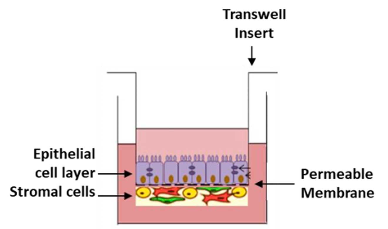
The question worth asking any platform: “Which cell in your model makes the collagen?” A model with no stromal compartment can report barrier and cytotoxicity. It cannot report an anti-fibrotic effect, because the fibrogenic cell is not in the well.
Immune-driven mechanisms
For compounds acting on T-cell, B-cell or macrophage activation rather than on the epithelium directly, two configurations are available:
- Conditioned medium — activated immune cells → supernatant → organoid
- Direct co-culture — immune cells cultured with the organoid
Evidence
The model separates compounds that look identical on paper
JAK inhibitors are not interchangeable. Across tofacitinib, upadacitinib and filgotinib, effects on fibrosis, permeability and inflammation diverge. Several reduce P1NP; barrier responses are not uniformly improved. IL-6 falls across the class, with the size of the effect depending on the donor. Anti-fibrotic and barrier effects separate — which is not visible in a model that reports only one axis.
A non-JAK compound matched JAK efficacy. FXR314 showed activity on barrier injury, inflammatory and fibrosis-associated readouts across both UC and CD models, broadly comparable to tofacitinib in the same system — establishing a non-JAK benchmark and demonstrating that mechanistically novel compounds can be positioned against the standard of care.
Clinically used anti-inflammatories are detected. Methotrexate produced a dose-dependent reduction in P1NP in both Crohn’s patient models, with statistically annotated reductions versus untreated control — confirming the fibrosis readout responds to drugs known to work in patients.
Publications
Dudum R, Payton O, Toohey C, et al. Digestive Disease Week 2025, Abstract No. Tu2040
Gervacio S, Dudum R, Aidnick H, et al. Digestive Disease Week 2025, Abstract No. Tu2035
Rachel Dudum, Mehrsa Mehrabi, Payton Olson, Cole Toohey, Haylee Aidnik, Caridad Rosette, Keith Murphy, Amar A. Sethi
Human 3D Liver Spheroid NAM for ADC DILI Risk Assessment
Mehrsa Mehrabi, Payton Olson, Cole Toohey, Rachel Dudum, Haylee Aidnik, Caridad Rosette, Keith Murhpy, Amar A. Sethi
Vivian (Xiao) Han, Haylee Aidnik, Sherwin Gervacio, Cole Toohey, Caridad Rosette, Keith Murphy, Amar A. Sethi
Caridad Rosette, Mehrsa Mehrabi, Rachel Dudum, Payton Olson, Cole Toohey, Haylee Aidnik, Philip Tan, Keith Murphy, Amar A. Sethi
Caridad Rosette, Vivian Xiao Han, Sherwin Gervacio, Cole Toohey, Haylee Aidnik, Keith Murphy, Amar A. Sethi
NAMkind™ Liver & Intestine Toxicity Prediction Across Small Molecules and Antibody Drug Conjugates
Amar A. Sethi, Mehrsa Mehrabi, Vivian Xiao Han, Rachel Dudum, Sherwin Gervacio, Payton Olson, Cole Toohey, Haylee Aidnik, Philip Tan, Caridad Rosette, and Keith Murphy
Sample Report
Does your MASH therapy deliver?
Brochure
How the service works
From intake to final report, the workflow is built for speed and clarity. You ship the compounds; we culture patient-derived transwells, run the repeat-dosing schedule, collect the endpoint panel, and translate the data into a recommendation.
Studies start the week the compounds and the purchase order arrive — there is no campaign queue. Interim status is visible on the customer portal throughout, and the final report is issued three weeks after the last treatment.
Standard service timeline
Day -3 onward Transwell seeding, differentiation and TEER baseline
Day 0 1st Dose
Weeks 3, 7, 10 Repeat dosing
Week 2 Harvest and endpoint collection
Weeks 3-5 Data Analysis
Week 5 Report and readout call
Flexible Study Design
- 14-day treatment period, 21-day option
- Apical or basolateral dosing
- 4+ concentrations per target
- Replicates set per endpoint
- 96-well transwell format
- UC and CD donors; healthy tissue comparator arm available
- Positive and negative controls on every plate
Included Assays
- TEER
- ATP
- Alamar Blue
- P1NP
- IL6
- Custom assays for additional fee are:
- immunohistochemistry for desired antigens
- Picrosirius Red (PSR) staining for collagen
- soluble fibrogenic biomarkers (ProC3, P3NP)
- RNAseq
Optional Assays
- Gene expression — RNA sequencing
- Extended cytokine panel — Luminex (IL-1β, MCP-1, TNF-α)
- Permeability to fluorescent tracers and macromolecules
- Immune co-culture configuration
- Multi-donor arms for variability assessment
- Reference compounds on every plate
- Positive: tofacitinib, upadacitinib
- Additional: methotrexate, prednisolone
- Negative: vehicle and untreated diseased control
Data Package
- Dose-response curves per endpoint, mean ± 95% CI
- Serial TEER traces across the treatment period
- Per-donor breakdown, not just the pooled mean
- Comprehensive tabular report and raw instrument files
- High-resolution microscopy images
- Interpreted slide deck and a scientist on the readout call

Real patient tissue, clinically annotated

Three disease axes, one well

Donor variability you can see

Clinically anchored on every plate
Getting started
Bring us the compound you are unsure about
The fastest way to evaluate the platform is to run one target you already have data on. If the model reproduces what you know, you can trust it on what you do not.
01 — Send the target and the question Compound class, indication, and what decision hangs on the result.
02 — Study design in 48 hours A written design and quote, reviewed with a VivoSim scientist.
03 — Pilot runs the week the PO lands No campaign queue. Interim status visible on the customer portal.
